Corneal Conditions
Pterygium Surgery in Camberley and Ascot
Specialist assessment and surgical removal of pterygium with Miss Hala Ali at Parkside Frimley, Camberley, and Parkside Heatherwood, Ascot.
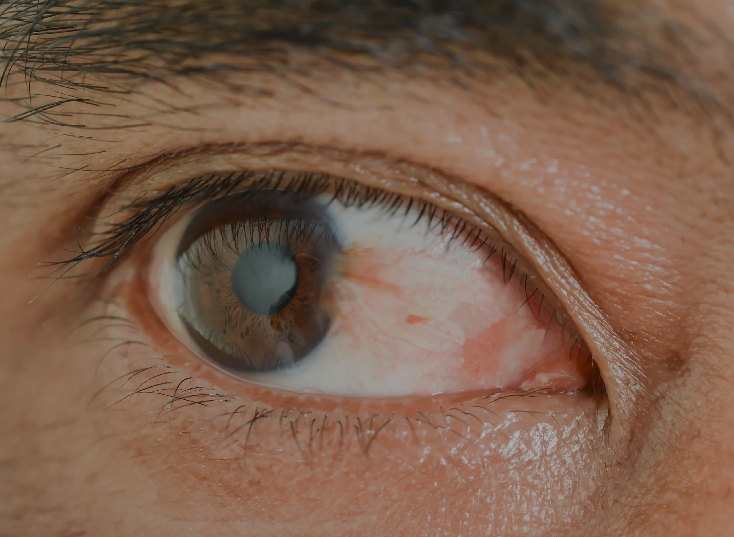
A pterygium is a benign, wedge-shaped growth of conjunctival tissue that extends from the white of the eye onto the cornea. Also known informally as "surfer's eye", it is associated with prolonged UV exposure and is more common in people who spend significant time outdoors. While small pterygia can be monitored, those causing persistent symptoms or threatening vision are treated by surgical excision. Miss Hala Ali uses the conjunctival autograft technique, which uses the patient's own tissue to cover the excision site and substantially reduces the risk of recurrence.
What is a pterygium?
A pterygium is a non-cancerous growth of the conjunctiva. This is the thin, transparent membrane covering the white of the eye. It typically arises from the inner corner of the eye and grows horizontally across the cornea, the clear front surface of the eye. In some cases it can arise from the outer side.
The growth is usually triangular or wing-shaped and may be pink, fleshy, or slightly raised. It is distinct from a pinguecula, which is a similar yellowish deposit that remains confined to the conjunctiva and does not extend onto the cornea.
Pterygium is not a malignant condition and does not spread to other parts of the body. However, if left untreated and allowed to grow towards the centre of the cornea, it can cause new astigmatism and reduce the vision.
Pterygium vs pinguecula
Pterygium
Grows from the conjunctiva onto the cornea. Can affect vision and cause astigmatism. May require surgical removal.
Pinguecula
Remains on the conjunctiva only. Does not extend onto the cornea. Usually managed conservatively with lubricating drops.
Symptoms of pterygium
Symptoms vary depending on the size and rate of growth of the pterygium. Some people have no symptoms at all, while others experience significant discomfort.
Persistent redness
The affected area of the eye appears chronically red or inflamed, even without infection.
Gritty or foreign body sensation
A feeling that something is in the eye, caused by the raised tissue on the ocular surface.
Dryness and irritation
The pterygium disrupts the tear film, leading to dryness and discomfort, particularly in wind or dry environments.
Blurred or distorted vision
As the pterygium grows towards the corneal centre, it can induce astigmatism and reduce visual clarity.
Visible fleshy growth
A triangular or wedge-shaped pinkish-white growth visible on the white of the eye, usually on the nasal side.
Difficulty with contact lenses
The raised tissue can make contact lens wear uncomfortable or impossible.
What causes pterygium?
The exact cause of pterygium is not fully understood, but several environmental and genetic factors are consistently associated with its development.
UV light exposure
Prolonged exposure to ultraviolet radiation is the most consistently identified risk factor. Pterygium is more common in people who spend significant time outdoors, particularly in sunny or high-altitude environments.
Dry and dusty environments
Chronic exposure to wind, dust, and low humidity irritates the conjunctiva and is associated with pterygium development and growth.
Geographic latitude
Pterygium is more prevalent in populations living closer to the equator, consistent with the role of UV exposure.
Genetic predisposition
A family history of pterygium may increase susceptibility, suggesting a genetic component alongside environmental triggers.
Diagnosis
Pterygium is diagnosed by clinical examination. During your consultation, Miss Hala Ali will:
- Examine the eye using a slit lamp biomicroscope to assess the size, extent, and vascularity of the pterygium
- Measure visual acuity and assess whether the pterygium is affecting vision
- Perform corneal topography to quantify any induced astigmatism
- Assess the rate of growth by reviewing any previous photographs or records where available
- Distinguish pterygium from other conditions such as pinguecula, conjunctival intraepithelial neoplasia, or limbal dermoid
The findings will be discussed with you in full, and a management plan — whether monitoring, conservative treatment, or surgery — will be agreed based on your symptoms and clinical findings.
Treatment options
The appropriate treatment depends on the size of the pterygium, the severity of symptoms, and whether it is affecting vision or corneal shape.
Conservative management
For small or stable pterygia that are not significantly affecting vision or comfort, conservative management may be appropriate:
- Preservative-free lubricating eye drops to relieve dryness and irritation
- UV-protective sunglasses to slow progression
- Short courses of topical anti-inflammatory drops during flare-ups
- Regular monitoring to detect growth towards the visual axis
Surgical excision with conjunctival autograft
Preferred technique for recurrence prevention
Surgery is recommended when the pterygium is symptomatic, growing, inducing astigmatism, or approaching the visual axis. The procedure involves:
- 1Local anaesthetic is applied
- 2The pterygium is carefully dissected from the cornea and sclera under the operating microscope
- 3A small free graft of conjunctival tissue is harvested from the upper part of the same eye
- 4The graft is secured over the excision site using tissue adhesive or fine sutures
- 5The procedure takes approximately 30 to 45 minutes as a day case
Who is suitable for pterygium surgery?
Surgery is typically considered when one or more of the following apply:
Significant or persistent symptoms
Redness, irritation, or foreign body sensation that is not adequately controlled with lubricating drops.
Growth towards the visual axis
The pterygium is encroaching on the central cornea and poses a risk to visual acuity.
Induced astigmatism
Corneal topography shows that the pterygium is distorting the corneal surface and affecting vision.
Pre-operative planning for cataract or refractive surgery
A pterygium should be removed before cataract or refractive lens surgery to allow accurate biometry and corneal measurements.
Suitability is determined at consultation. Miss Hala Ali will discuss the risks and benefits of surgery in the context of your individual findings and preferences.
Clinic locations
Miss Hala Ali holds private clinics at two locations in Surrey and Berkshire, offering convenient access for patients across the region.
Parkside Frimley — Camberley
Monday evenings
Frimley Road, Camberley, Surrey, GU16 7QJ
Parkside Heatherwood — Ascot
Thursday and Friday
Brook Avenue, Ascot, Berkshire, SL5 7GB
Consultation fees
An initial consultation with Miss Hala Ali includes a full clinical assessment, slit lamp examination, and corneal topography where indicated. Surgical fees are discussed and confirmed at consultation.
New consultation
£250
Follow-up consultation
£200
Miss Hala Ali accepts most major private medical insurers including Bupa, AXA Health, Vitality, Allianz, WPA, Aviva, Cigna, Aetna, The Exeter, Healix Health, and General & Medical. Please check your policy for ophthalmology cover before booking.
View full fees informationFrequently asked questions
What is a pterygium?
A pterygium is a benign, fleshy growth of conjunctival tissue that extends from the white of the eye onto the cornea. It is not a cancer and does not spread beyond the eye, but it can cause redness, irritation, and visual disturbance if it grows towards the centre of the cornea.
Does a pterygium always need surgery?
Not always. Small pterygia that are not causing significant symptoms or threatening vision can be monitored and managed with lubricating drops and sun protection. Surgery is recommended when the pterygium is causing persistent symptoms, is growing towards the visual axis, or is inducing significant astigmatism.
What is a conjunctival autograft and why is it used?
A conjunctival autograft involves taking a small piece of the patient's own conjunctival tissue from the upper part of the eye and using it to cover the area where the pterygium was removed. This technique significantly reduces the risk of recurrence compared with bare sclera excision alone.
Will my pterygium come back after surgery?
Recurrence is possible but is substantially reduced by the conjunctival autograft technique. Protecting the eyes from UV light and dust after surgery also helps reduce the risk of recurrence.
How long is recovery after pterygium surgery?
Most patients experience some redness, grittiness, and mild discomfort for one to two weeks. Vision is usually functional within a few days, though the eye may remain red for several weeks as it heals. Topical drops are prescribed for several weeks post-operatively.
Is pterygium surgery performed under general or local anaesthetic?
Pterygium surgery is performed under local anaesthetic as a day-case procedure. You will be awake but the eye is completely numb. General anaesthetic is not required.
Where does Miss Hala Ali perform pterygium surgery?
Miss Hala Ali performs pterygium surgery at Parkside Frimley in Camberley, Surrey, and Parkside Heatherwood in Ascot, Berkshire.
Part of the Corneal Conditions service
Miss Hala Ali also manages keratoconus, corneal dystrophies, corneal infections, and other corneal conditions.
Concerned about a growth on your eye?
Book a private consultation with Miss Hala Ali in Camberley or Ascot for a specialist assessment and personalised management plan.